Learn · General Surgery · Intermediate
Small bowel obstruction
The abdominal X-ray from a Code Status case, and why the urgent question is whether the bowel is losing its blood supply.
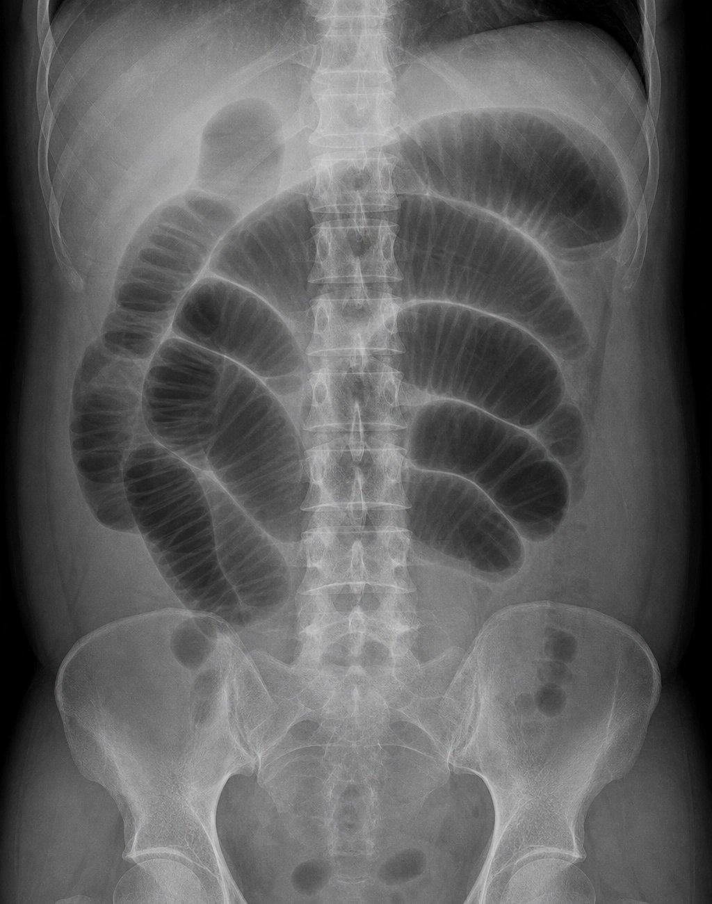
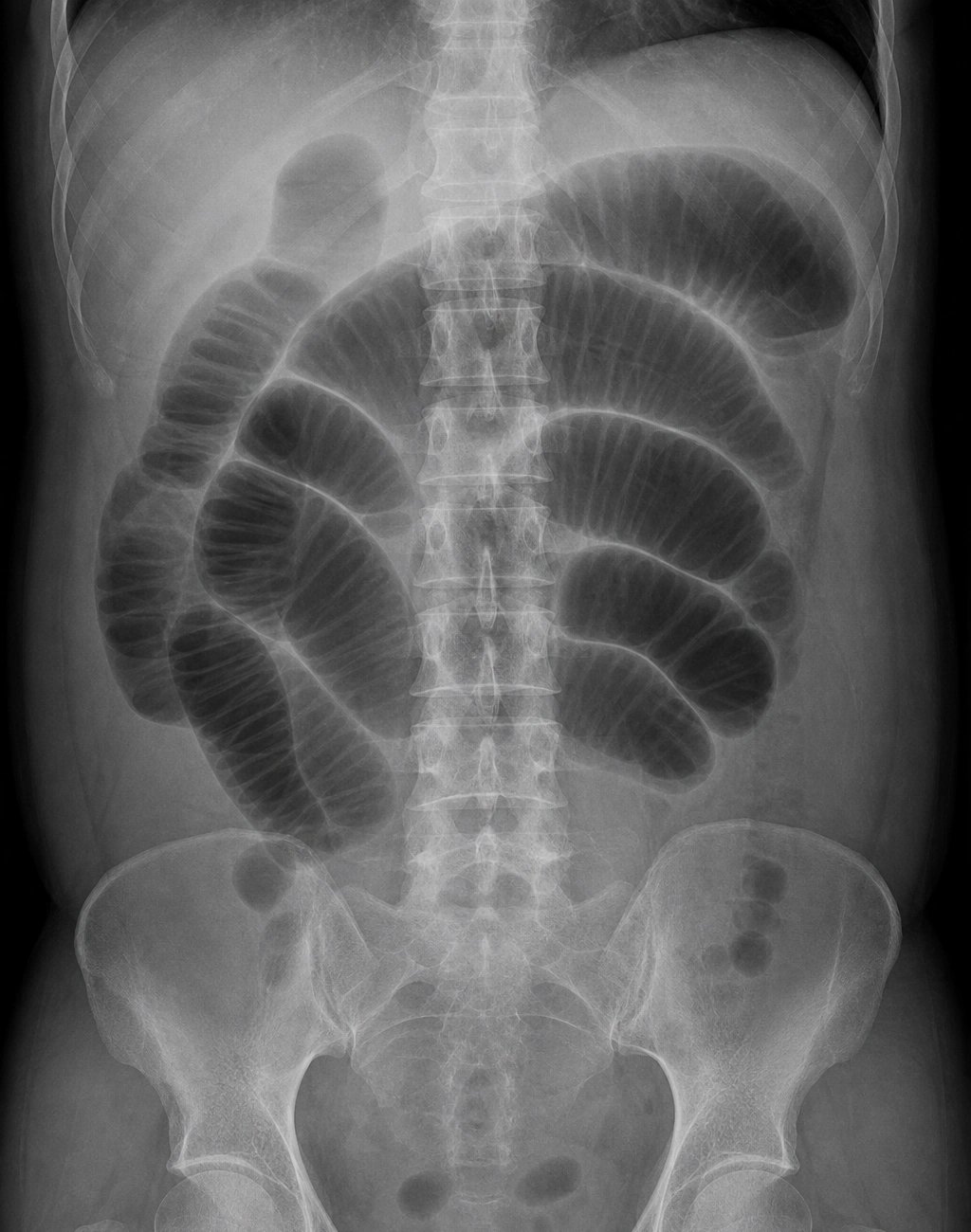
Dilated loops of small bowel fill the middle of the abdomen.
Linda Park’s abdominal X-ray, from her case. Open it full size
Reading this X-ray
Start in the middle of the abdomen. Several gas-filled loops of small bowel, stacked on both sides of the spine, are wider than normal. Thin white folds, the folds of the small bowel lining, cross the full width of each loop. Together, these findings suggest small bowel obstruction.
Air-fluid levels, flat lines where gas sits on top of fluid, are not visible on this film, which was taken lying down. They show on a film taken upright, so their absence here does not rule out obstruction. A plain film can also occasionally show free air in the abdomen.
An X-ray cannot reliably show where the blockage is, what caused it or whether the bowel is losing its blood supply. CT of the abdomen and pelvis, with contrast through a vein, is the usual next study when obstruction is suspected. It shows where dilated bowel changes to collapsed bowel, suggests the cause and looks for complications.
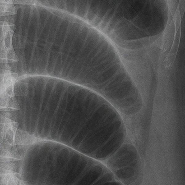
Dilated small bowel loops, enlarged
In the emergency department
Recognize the obstruction, find the cause, then protect the bowel.
On arrival
Recognize the pattern
Cramping abdominal pain, vomiting and a swollen belly suggest bowel obstruction. The pain often comes in waves as the bowel contracts against the blockage. Passing no stool or gas raises concern for a high-grade obstruction, but recent bowel movements do not rule out a partial one. Linda, 62, has had pain in waves since yesterday, has vomited four times and has passed no gas since last night.
History and examination
Look for the cause
Previous abdominal surgery is the strongest clue to adhesions, bands of scar tissue inside the abdomen that can block the bowel years or decades later. Linda had an open hysterectomy fifteen years ago. Examine for hernias, including both groins and every old scar. With no previous surgery, a hernia or a cancer moves up the list.
At every examination
Recognize threatened bowel
The danger is strangulation: a blockage that cuts off the bowel’s blood supply (ischemia). Watch for pain that becomes constant or more severe, guarding, rebound tenderness, a fever, a rising heart rate or a falling blood pressure. These can develop over hours, so examine again and again. A normal lactate does not rule out early ischemia.
Imaging
Look beyond the blockage
CT shows where the blockage is and looks for complications. Reduced enhancement of the bowel wall, meaning it takes up less contrast, raises concern for ischemia, but normal enhancement does not guarantee healthy bowel. A closed loop, a segment blocked at two points, is especially dangerous. Gas in the bowel wall or in the portal veins, or free air in the abdomen, can signal advanced injury or a hole in the bowel (perforation). Read these findings with the examination and the course over time.
Treatment
Replace losses and decompress
Nothing by mouth. Give isotonic fluid through a vein to replace what vomiting has lost and what is trapped in the swollen bowel. Check kidney function and electrolytes, and correct them: vomiting loses stomach acid, so chloride and potassium fall. Treat pain and nausea, but avoid drugs that make the bowel contract, such as metoclopramide, against a mechanical block. A nasogastric tube, passed through the nose into the stomach, decompresses the bowel and helps most with ongoing vomiting or a very swollen belly.
Then decide
Involve surgery early
Involve the surgeons early for any suspected mechanical blockage. A rigid or guarding abdomen, suspected ischemia or strangulation, perforation, a closed loop or a patient who is getting worse needs urgent surgical review. A stable patient with an adhesive blockage and no sign of threatened bowel may settle without an operation: admission, nothing by mouth, fluids through a vein, decompression and repeated examinations. Water-soluble contrast given down the nasogastric tube can help predict who will settle. A trial of about 72 hours is reasonable in selected stable patients, but worsening findings call for action sooner. Antibiotics are not routine: give them when ischemia or perforation is suspected.
References (6)
- Bologna Guidelines for Diagnosis and Management of Adhesive Small Bowel Obstruction (ASBO): 2017 Update. World Journal of Emergency Surgery, 2018.
- Evaluation and Management of Small-Bowel Obstruction: An Eastern Association for the Surgery of Trauma Practice Management Guideline. Journal of Trauma and Acute Care Surgery, 2012.
- ACR Appropriateness Criteria Suspected Small-Bowel Obstruction. Journal of the American College of Radiology, 2020.
- Acute Mesenteric Ischemia: Updated Guidelines of the World Society of Emergency Surgery. World Journal of Emergency Surgery, 2022.
- Value of CT Findings to Predict Surgical Ischemia in Small Bowel Obstruction: A Systematic Review and Meta-analysis. European Radiology, 2015.
- Comparing Outcomes of Nonoperative Treatment for Adhesive Small Bowel Obstruction With and Without Antibiotics. Journal of Infection and Chemotherapy, 2021.
In the app, Linda’s Attending’s Notes go deeper: the electrolytes, the nasogastric tube, the treatment without an operation and the evidence, with 10 sources linked to the originals.
Written by Karim Ali, MD, board-certified emergency physician. Reviewed November 2026.
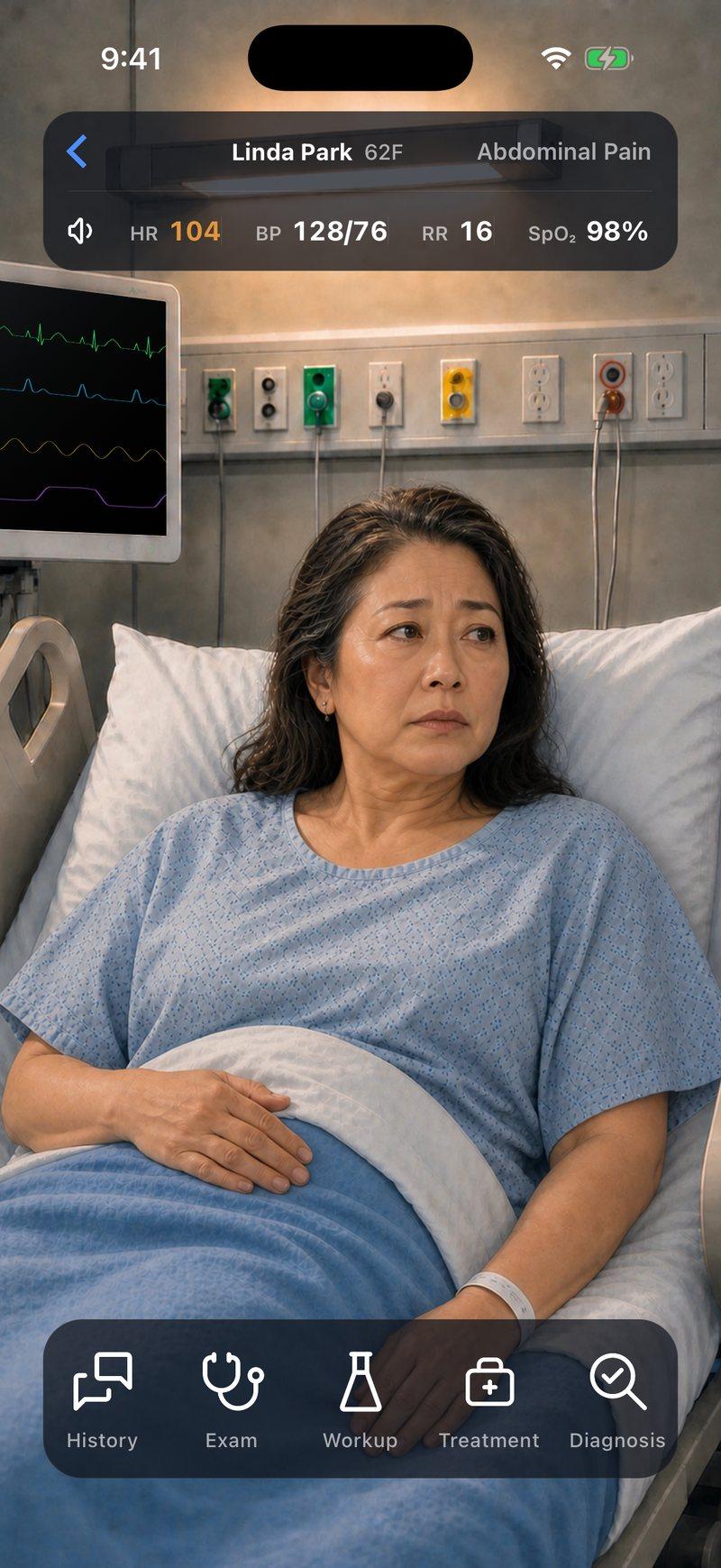
Now make the call yourself
Linda Park, 62, has abdominal pain that comes in waves. She has vomited four times and has passed no gas since last night. Take her history, examine her, read her imaging and decide whether she needs surgery.


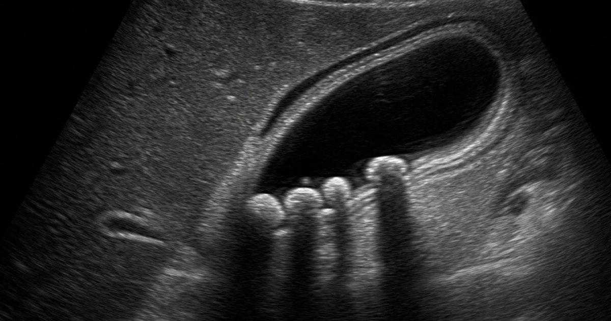
Linda is a fictional patient, and this page is a summary for learning. Code Status is for training only and is not intended for real-world patient care, diagnosis, treatment, or clinical decision-making.