Learn · Nephrology · Advanced
Hyperkalemia
The ECG from a Code Status case, and the treatment that buys time until dialysis.
Written by Karim Ali, MD, board-certified emergency physician. Reviewed November 2026.
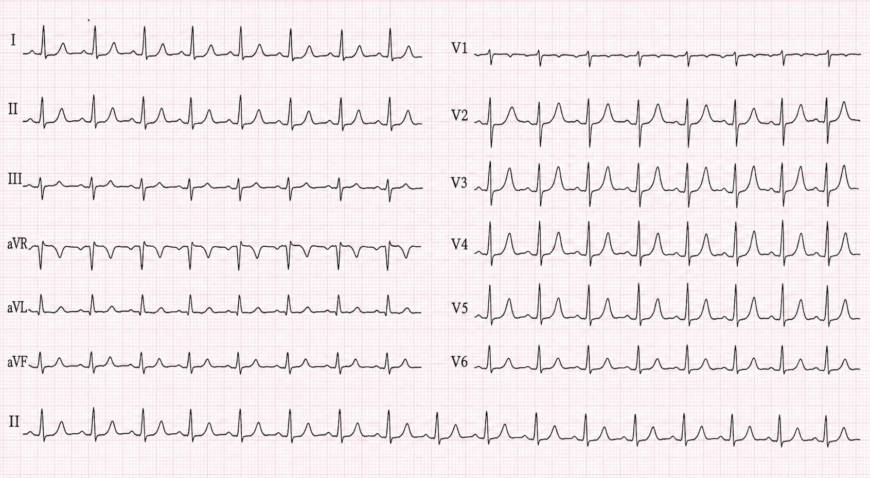
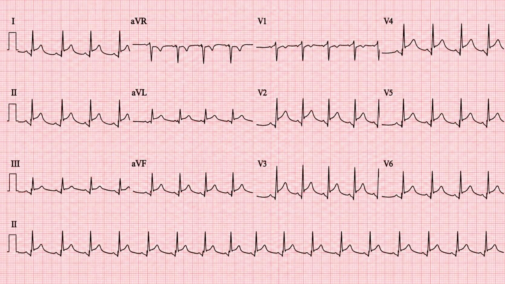
Tall, pointed T waves, clearest in the chest leads V2 to V5. This is Walter’s ECG after his pulse returned, and his potassium was still high.
Walter Higgins’s ECG, from his case. Open it full size
Reading this ECG
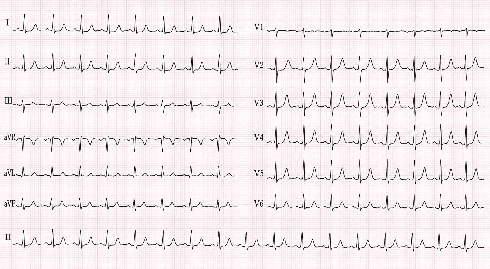
Look at the T waves. Here, in V3, the T wave is about 7 mm tall at standard calibration, almost as tall as the R wave, with a pointed, symmetrical peak. That tall, tented shape is the peaked T wave of high potassium.
Tall T waves are not always potassium, though. The hyperacute T waves of an early STEMI, early repolarization and left ventricular hypertrophy can all make them tall, so read them with the story and the potassium, and repeat the ECG when you are unsure.
As potassium rises, other changes can follow: smaller P waves, a longer PR interval, a widening QRS and, at the extreme, a sine-wave pattern. They do not reliably appear in that order. Walter’s complexes were wide during his arrest. In this tracing, taken after his pulse returned, the QRS is narrow but the T waves are still peaked.
Two traps. A normal-looking ECG does not rule out severe hyperkalemia: the ECG is an insensitive test for high potassium, and severe hyperkalemia can come with no ECG changes at all. And a better ECG does not mean the potassium is down: after treatment, Walter’s potassium was still high.
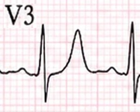
V3, enlarged
In the emergency department
Protect the heart, shift the potassium, remove it. These are the steps for Walter, in cardiac arrest. Outside an arrest the same three jobs apply, but whether calcium is needed and how fast each step happens depend on the potassium level, the ECG and local protocol.
Suspect it
Think of it before the result is back
Kidney failure or missed dialysis, medicines or other causes that raise potassium or stop the kidneys clearing it, a slow heart rate or heart block before a collapse, or a widening QRS should make you think of high potassium. Walter had missed his last two dialysis sessions. In a cardiac arrest where it is strongly suspected, start treatment alongside CPR without waiting for the potassium result. His came back at 7.9 mmol/L. Evidence that calcium, insulin with glucose or bicarbonate improves survival from a hyperkalemic arrest is uncertain, and none of them replaces good CPR.
First
Protect the heart with calcium
Intravenous calcium counters potassium’s effect on the heart within minutes. It does not lower potassium, and its protection is temporary. Calcium gluconate is gentler on a peripheral vein than calcium chloride, but do not delay urgent treatment to place a central line. In a cardiac arrest, calcium is not routine. It may be given when high potassium is strongly suspected, as it was for Walter.
Next
Shift the potassium into cells
Insulin with glucose moves potassium into cells, but only for a while. Check glucose repeatedly for at least six hours, because insulin can cause low blood sugar hours later, especially in kidney failure. Once a pulse has returned, inhaled albuterol (salbutamol) can add to the effect, but never on its own.
At the same time
Arrange dialysis early
Calcium and insulin only buy time. Dialysis removes the potassium, so call for the team, access and equipment early. Sodium bicarbonate is not a routine potassium-lowering treatment. It may be considered when severe metabolic acidosis is part of the problem, as it was for Walter, but evidence of benefit in a hyperkalemic arrest is uncertain.
When the result comes back
Read the number, and make sure it is real
There is no single agreed grading of hyperkalemia. It is commonly defined as 5.5 mmol/L or above, and the KDIGO kidney conference recommends cardiac monitoring and a 12-lead ECG above 6.0. That is not a floor: ECG changes, symptoms or a fast-rising level call for monitoring and urgent treatment at any level. A high result can also be false: blood damaged (hemolyzed) in the draw, a long tourniquet or fist clenching, a clotted sample, or a very high white cell or platelet count. Suspect this when the result does not fit the patient, and repeat it, but never let a repeat delay treatment for someone like Walter.
Ongoing care
Watch for the rebound
Potassium can climb again as the shifting treatment wears off. Recheck it at about 1, 2, 4 and 6 hours, give calcium again if the ECG changes persist or come back, and keep checking glucose. A pulse and a better ECG do not mean the danger has passed.
References (6)
- Part 10: Adult and Pediatric Special Circumstances of Resuscitation: 2025 AHA Guidelines for CPR and ECC. Circulation, 2025.
- European Resuscitation Council Guidelines 2025 Special Circumstances in Resuscitation. Resuscitation, 2025.
- UK Kidney Association Clinical Practice Guideline: Management of Hyperkalaemia in Adults. UK Kidney Association, 2023.
- Acute hyperkalemia in the emergency department: a summary from a Kidney Disease: Improving Global Outcomes conference. European Journal of Emergency Medicine, 2020.
- The prominent T wave: electrocardiographic differential diagnosis. American Journal of Emergency Medicine, 2002.
- Hypoglycaemia due to insulin therapy for the management of hyperkalaemia in hospitalised adults: A scoping review. PLOS ONE, 2022.
In the app, Walter’s Attending’s Notes go deeper: doses, pitfalls and the evidence, with 10 sources linked to the originals.
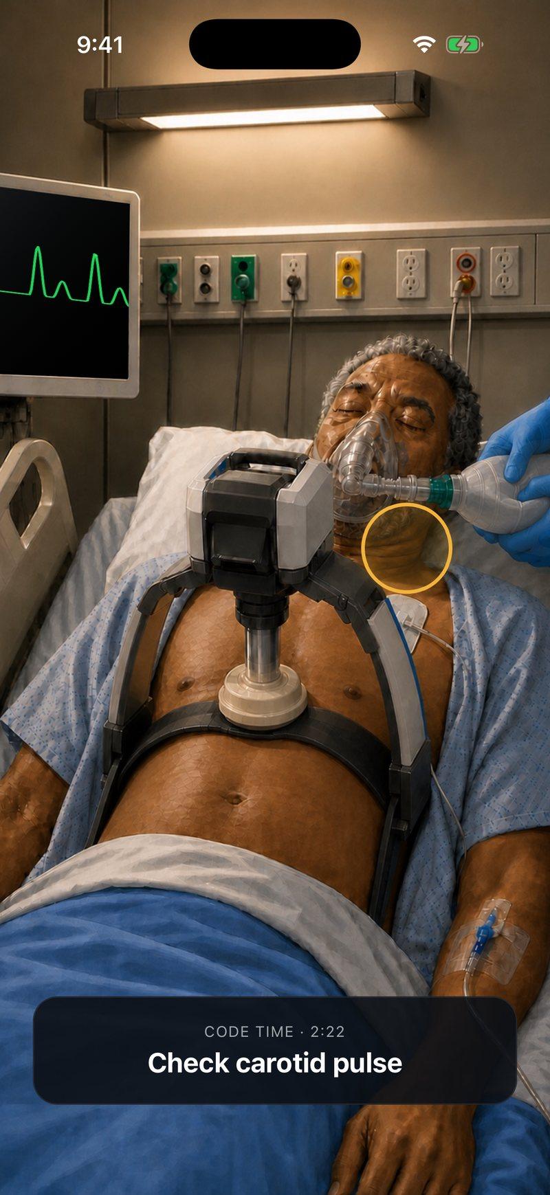
Now make the call yourself
Walter Higgins, 70, arrives in cardiac arrest with CPR underway. He has kidney failure and missed his last two dialysis sessions. Run the resuscitation, find the cause and decide what happens next.


Walter is a fictional patient, and this page is a summary for learning. Code Status is for training only and is not intended for real-world patient care, diagnosis, treatment, or clinical decision-making.