Learn · Orthopedics · Intermediate
Anterior shoulder dislocation
The X-ray from a Code Status case, and how to put the shoulder back safely.
Written by Karim Ali, MD, board-certified emergency physician. Reviewed November 2026.
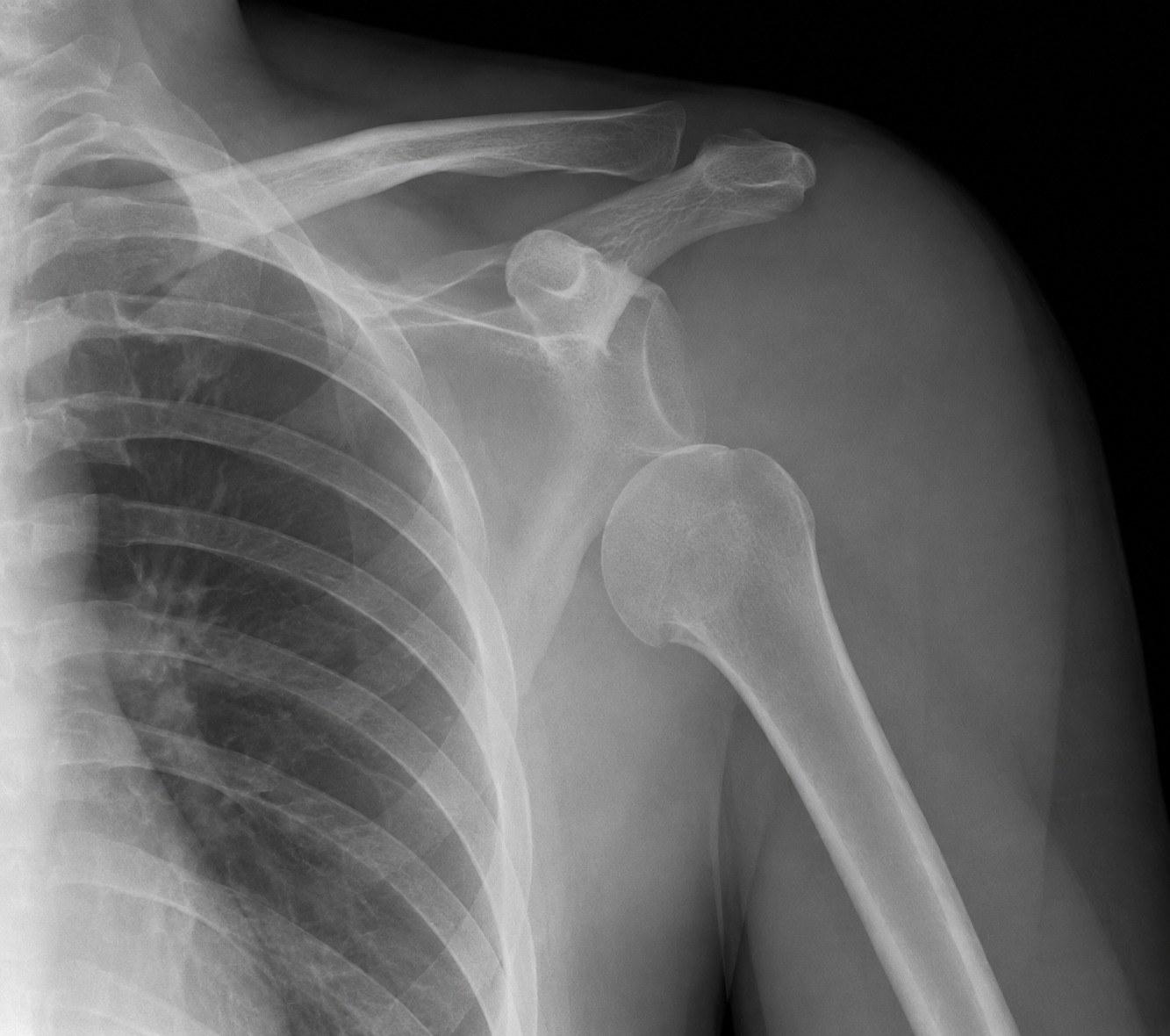
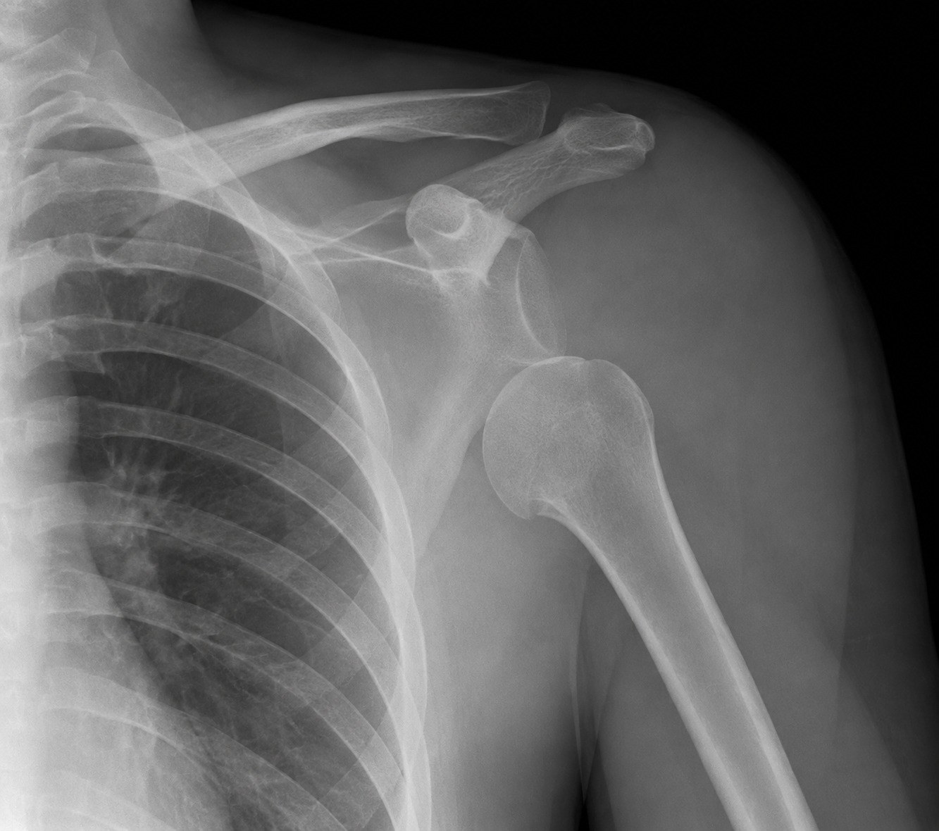
The ball of the upper arm bone sits below the socket instead of in it. This is Jordan’s left shoulder, seen from the front.
Jordan Reyes’s shoulder X-ray, from his case. Open it full size
Reading this X-ray
Find the ball and the socket. On a front view of a normal shoulder, the round head of the humerus sits against the glenoid, the shallow socket of the shoulder blade, like a golf ball on a tee.
Here the socket is empty. The humeral head sits below it, where it came to rest after being pushed forward out of the joint. Before anyone puts it back, trace the head, the neck of the humerus and the rim of the socket for a fracture.
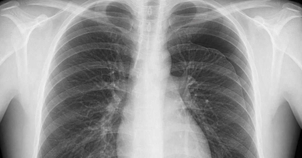
After reduction, the head sits back against the socket, centered on it. A film that is not centered means the joint is not back in, whatever the maneuver felt like.
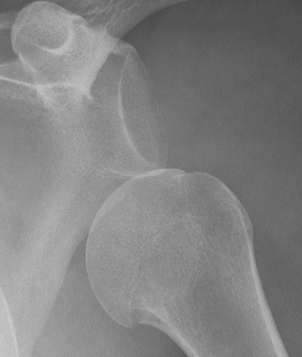
Before reduction
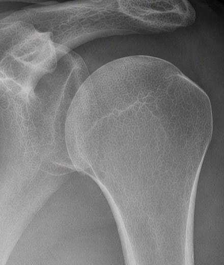
After reduction
In the emergency department
From the first look to the sling, in order.
On arrival
Recognize the dislocation
Jordan, 26, fell while rock climbing. His arm was caught and pulled backward and upward: forced abduction and external rotation, the classic setup. The shoulder looks squared off, the arm is held slightly away from the body, and he guards against any movement.
Before you touch it
Check the nerves and the pulse
The axillary nerve wraps around the surgical neck of the humerus, right in the path of the displaced head. Test light touch over the outer shoulder and the deltoid contracting with gentle effort, then movement and sensation further down the arm, the radial pulse and capillary refill. A normal pulse alone does not make the exam normal. Write it down before any sedation or manipulation, so you can compare afterward.
Before reduction
Look for a fracture first
Think of a fracture in older patients, after a high-energy injury, with bony tenderness away from the joint, or when the arm position does not fit the pattern. A greater tuberosity fracture often goes back with the joint, but a fracture through the neck of the humerus can turn a bedside reduction into a surgical problem. When a fracture is plausible, image before reducing.
Pain control
Relax the shoulder
A relaxed shoulder is easier and safer to reduce, and not everyone needs deep sedation. A cooperative patient with a first-time dislocation often does well with lidocaine injected into the joint, or a regional block, and a gentle technique. Save procedural sedation for heavy guarding, significant anxiety or a lighter approach that has failed, and set up a monitor, continuous capnography and a second clinician for the airway first.
The reduction
Reduce it gently
With the patient lying down, the elbow bent to 90 degrees and the arm at the side, slowly rotate the forearm outward, pausing at each point of resistance until the spasm relaxes. Scapular manipulation is another gentle option: it turns the socket to meet the ball. If it will not go back, do not pull harder. Improve relaxation, positioning or technique, and think again about a fracture. Stop if each try needs more force or a new nerve or pulse change appears, then re-image and involve orthopedics.
Afterward
Prove it worked
On the film after reduction, the head should sit centered in the socket. If it does not, the joint is not back in, whatever the maneuver felt like. Read that film for a fracture too. Repeat the nerve and pulse exam and compare it with the first one: new weakness, numbness or unexpected pain needs another look and orthopedics before discharge. Then a sling, follow-up, and a clear warning that it can happen again, because the risk is substantial in young, active patients.
References (6)
- ACR Appropriateness Criteria: Acute Shoulder Pain, 2024 Update. Journal of the American College of Radiology, 2025.
- An Umbrella Review of Systematic Reviews and Meta-analyses for Assessment and Treatment of Acute Shoulder Dislocation. American Journal of Emergency Medicine, 2025.
- A Systematic Review With Pairwise and Network Meta-analysis of Closed Reduction Methods for Anterior Shoulder Dislocation. Annals of Emergency Medicine, 2023.
- Intra-articular Lidocaine Versus Intravenous Sedation for Closed Reduction of Acute Anterior Shoulder Dislocation in the Emergency Department: A Systematic Review and Meta-analysis. Canadian Journal of Emergency Medicine, 2022.
- ACEP Clinical Policy: Procedural Sedation and Analgesia in the Emergency Department. Annals of Emergency Medicine, 2014.
- Shoulder Stabilization Versus Immobilization for First-Time Anterior Shoulder Dislocation: A Systematic Review and Meta-analysis of Level 1 Randomized Controlled Trials. American Journal of Sports Medicine, 2023.
In the app, Jordan’s Attending’s Notes go deeper: the procedure step by step, pitfalls and the evidence, with 10 sources linked to the originals.
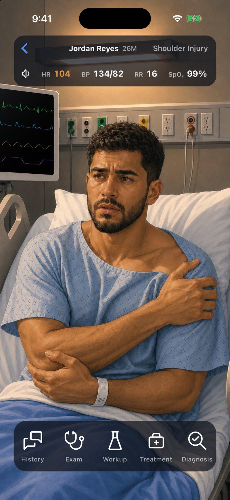
Now make the call yourself
Jordan Reyes, 26, falls while rock climbing and arrives with severe pain in his left shoulder. Examine him, read his X-ray, control his pain and decide how to put it back.


Jordan is a fictional patient, and this page is a summary for learning. Code Status is for training only and is not intended for real-world patient care, diagnosis, treatment, or clinical decision-making.