Learn · Pulmonology · Advanced
Spontaneous pneumothorax
The chest X-ray from a Code Status case, and why stability and symptoms, not size alone, decide what happens next.
Written by Karim Ali, MD, board-certified emergency physician. Reviewed November 2026.
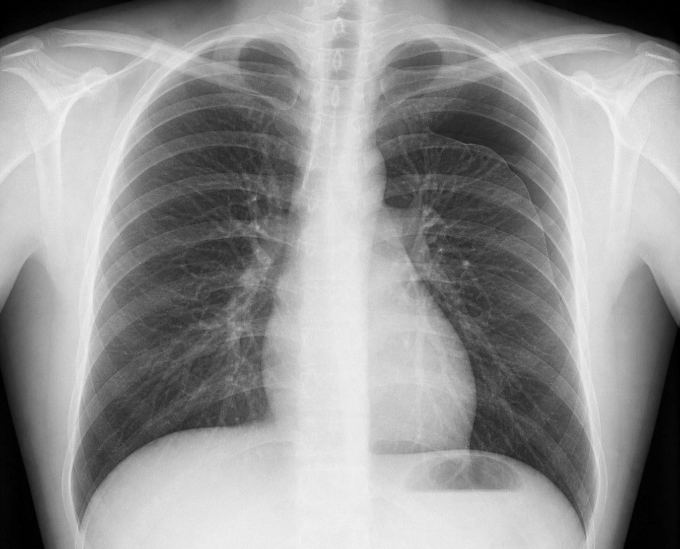
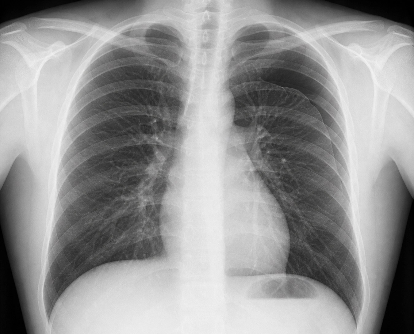
A thin white line at the top of the left lung, with no lung markings beyond it. On a chest X-ray, the patient’s left is on your right.
Mateo Alvarez’s chest X-ray, from his case. Open it full size
Reading this X-ray
Look at the top of each lung. Here, at the top of the left lung, a thin white line runs across the upper chest and curves down along its side. That is the pleural line, the edge of the lung where it has pulled away from the chest wall. Between the line and the ribs there are no lung markings, only air.
The heart and trachea sit in the middle.
At the bedside, ultrasound can help. Lung sliding, the two layers of pleura moving together with each breath, rules out a pneumothorax at that spot. Absent sliding is not enough on its own: stuck pleura, a lung that is not being ventilated or a held breath can remove it too. A lung point, where sliding appears and disappears at the edge of the air, strongly supports the diagnosis.
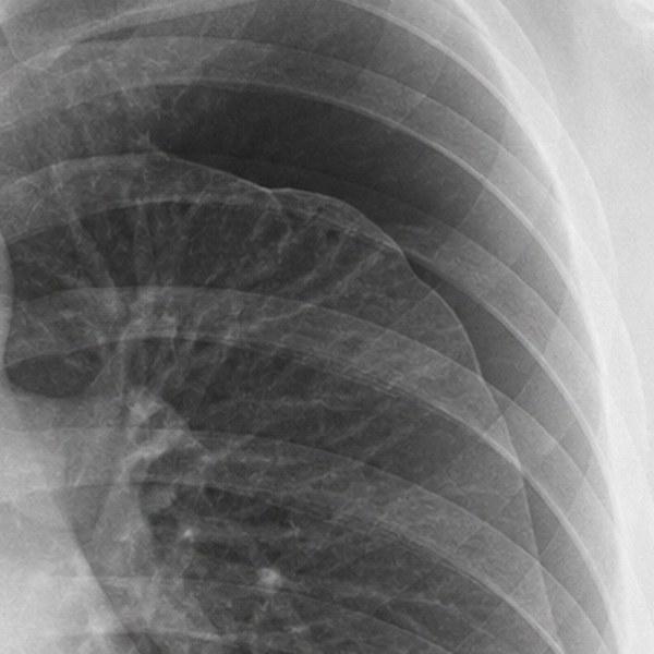
Left upper chest, enlarged
In the emergency department
Stability first, then symptoms, then size.
On arrival
Ask whether the patient is unstable
The first question is not how big the pneumothorax is. It is whether the patient is unstable. Mateo, 27, had sudden sharp left-sided chest pain at rest and was short of breath. His heart rate was 104 and his oxygen saturation 92% on room air, but his blood pressure was normal and he was speaking in full sentences.
If the patient is unstable
Tension is a bedside diagnosis
Air can build up in the pleural space faster than it escapes, as when a tear in the lung acts as a one-way valve. The rising pressure squeezes the veins returning blood to the heart, causing obstructive shock. A patient with a suspected pneumothorax and worsening breathing or circulation may need decompression at once. Do not wait for an X-ray, or for the trachea to shift, which is often absent. A film with the heart and trachea in the middle does not rule it out either. Decompression only buys time, so a chest tube follows promptly.
If the patient is stable
Primary or secondary
Primary spontaneous pneumothorax happens in a lung with no apparent disease, classically in tall, thin young people, often at rest. Secondary means there is lung disease underneath, most often emphysema. Those patients have less reserve, so the bar for drainage and admission is generally lower.
Then decide
Observe, aspirate or drain
Size alone does not decide. A stable primary pneumothorax with few symptoms may be observed, if the patient can be followed up and return quickly. Persistent symptoms or low oxygen call for removing the air: a small catheter or chest tube connected to a water seal, or, for a stable primary pneumothorax in some settings, simple aspiration. Oxygen treats low oxygen, but it never replaces removing the air. Mateo was breathless at rest with an oxygen saturation of 92%. For him, that meant oxygen and removing the air, rather than observation alone.
Placing a drain
Use the safe triangle
Confirm the side first. Go through the fourth or fifth intercostal space inside the safe triangle. Its borders are the lateral edge of pectoralis major in front, the front edge of latissimus dorsi behind and a line at the level of the fifth intercostal space below, and its point sits just below the armpit. Enter just above the rib below, away from the nerve and vessels under each rib, and never too low, where the liver, spleen and diaphragm sit. For air alone, a small soft catheter placed over a guidewire drains as well as a large tube in most patients.
After the drain
Read the drainage system
Water rising and falling with each breath (tidaling) suggests the tube is open to the chest. It also fades once the lung is fully up. Bubbling means air is leaving. If the water stops moving and the breathlessness comes back, check the patient and the tubing at once: a kink, a block, a loose connection or a displaced tube. Do not clamp the tube to troubleshoot, because that can recreate tension. The drain comes out when the leak has stopped and the lung has stayed up.
References (6)
- Joint ERS/EACTS/ESTS Clinical Practice Guidelines on Adults With Spontaneous Pneumothorax. European Respiratory Journal, 2024.
- British Thoracic Society Guideline for Pleural Disease. Thorax, 2023.
- British Thoracic Society Clinical Statement on Pleural Procedures. Thorax, 2023.
- Conservative Versus Interventional Treatment for Spontaneous Pneumothorax. New England Journal of Medicine, 2020.
- Observation, Aspiration, or Tube Thoracostomy for Primary Spontaneous Pneumothorax: A Systematic Review, Meta-analysis, and Cost-Utility Analysis. CHEST, 2023.
- A Systematic Review and Meta-analysis Comparing Pigtail Catheter and Chest Tube as the Initial Treatment for Pneumothorax. CHEST, 2018.
In the app, Mateo’s Attending’s Notes go deeper: the procedure step by step, pitfalls and the evidence, with 10 sources linked to the originals.

Now make the call yourself
Mateo Alvarez, 27, arrives with sudden sharp chest pain on the left and cannot take a full breath. Assess his stability, read his chest X-ray and decide what happens next.


Mateo is a fictional patient, and this page is a summary for learning. Code Status is for training only and is not intended for real-world patient care, diagnosis, treatment, or clinical decision-making.