Learn · Pulmonology · Intermediate
Community-acquired pneumonia
The chest X-ray from a Code Status case, and why breathing, circulation and mental state, not a score alone, decide where the patient is treated.
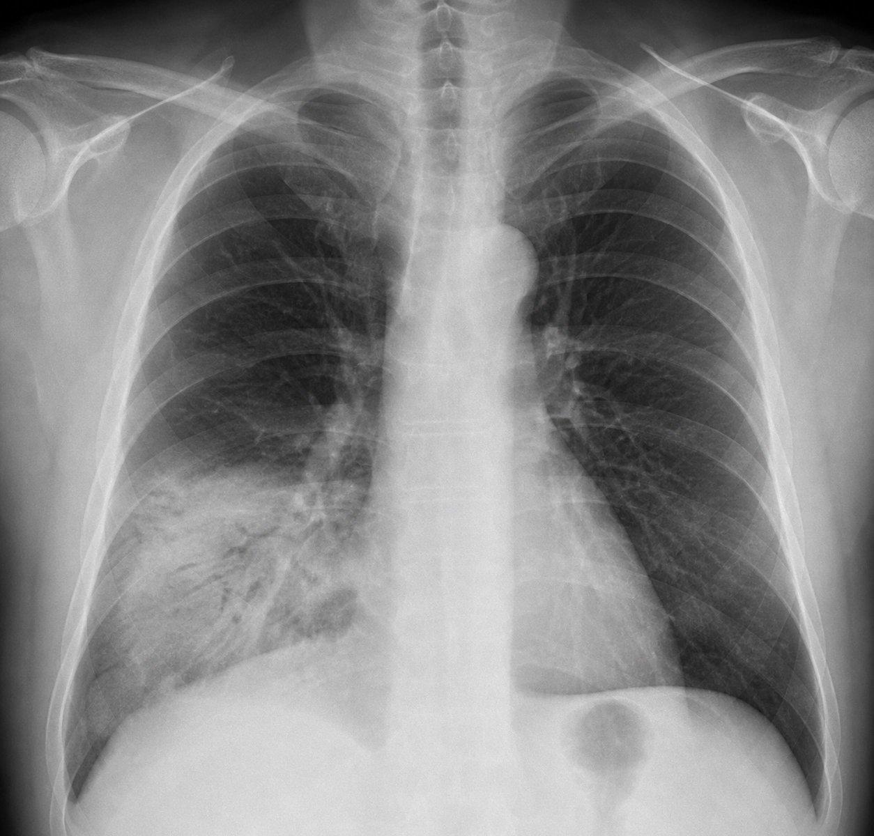
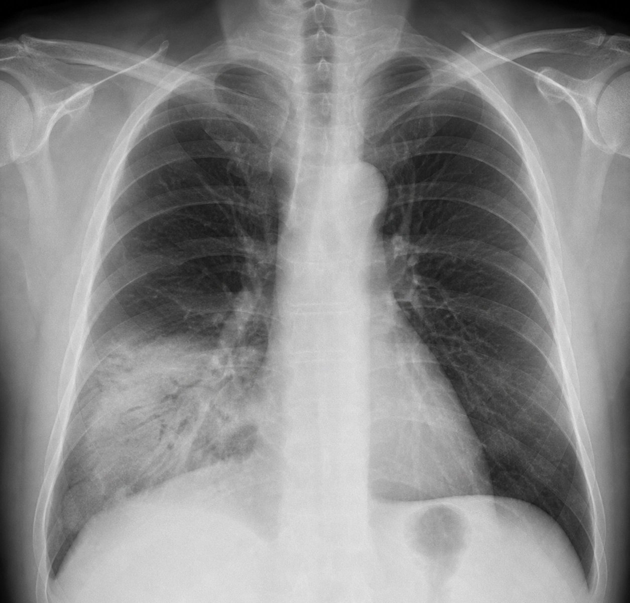
A white area at the base of the right lung, with dark branching airways running through it.
Daniel Kim’s chest X-ray, from his case. Open it full size
Reading this X-ray
Compare the lower part of each lung. Here, at the base of the right lung, a white area replaces the dark, air-filled lung seen above it and on the other side. That is consolidation: the air spaces have filled with fluid or cells.
Inside the white area, dark branching lines run through it. These are air bronchograms, airways that still hold air, outlined by the filled lung around them. They support a process in the air spaces, but they are not specific for infection. Read the film together with the fever and the cough.
Consolidation is not always infection. Collapsed lung (atelectasis), a pulmonary infarct, aspiration and a mass blocking an airway can look similar, and collapse pulls nearby structures toward the white area. A normal film very early does not rule out pneumonia when suspicion stays high. CT is for a suspected complication, a patient who is not improving or a genuinely open question, not for straightforward pneumonia.
At the bedside, lung ultrasound can show consolidation near the surface of the lung, without radiation: a solid-looking area under the pleura with bright air bronchograms inside it. It depends on the operator and cannot see disease that does not reach the pleura. Fluid on the lungs from heart failure typically shows B-lines on both sides instead of one focal area.
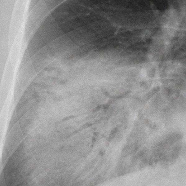
Right lung base, enlarged
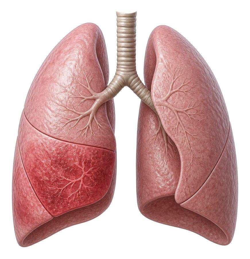
What is happening
The drawing shows where the infection sits: the lower lobe of the right lung is filled in, while the rest of both lungs still holds air.
In the emergency department
Recognize it, judge severity, treat promptly, then decide where.
On arrival
Recognize the pattern
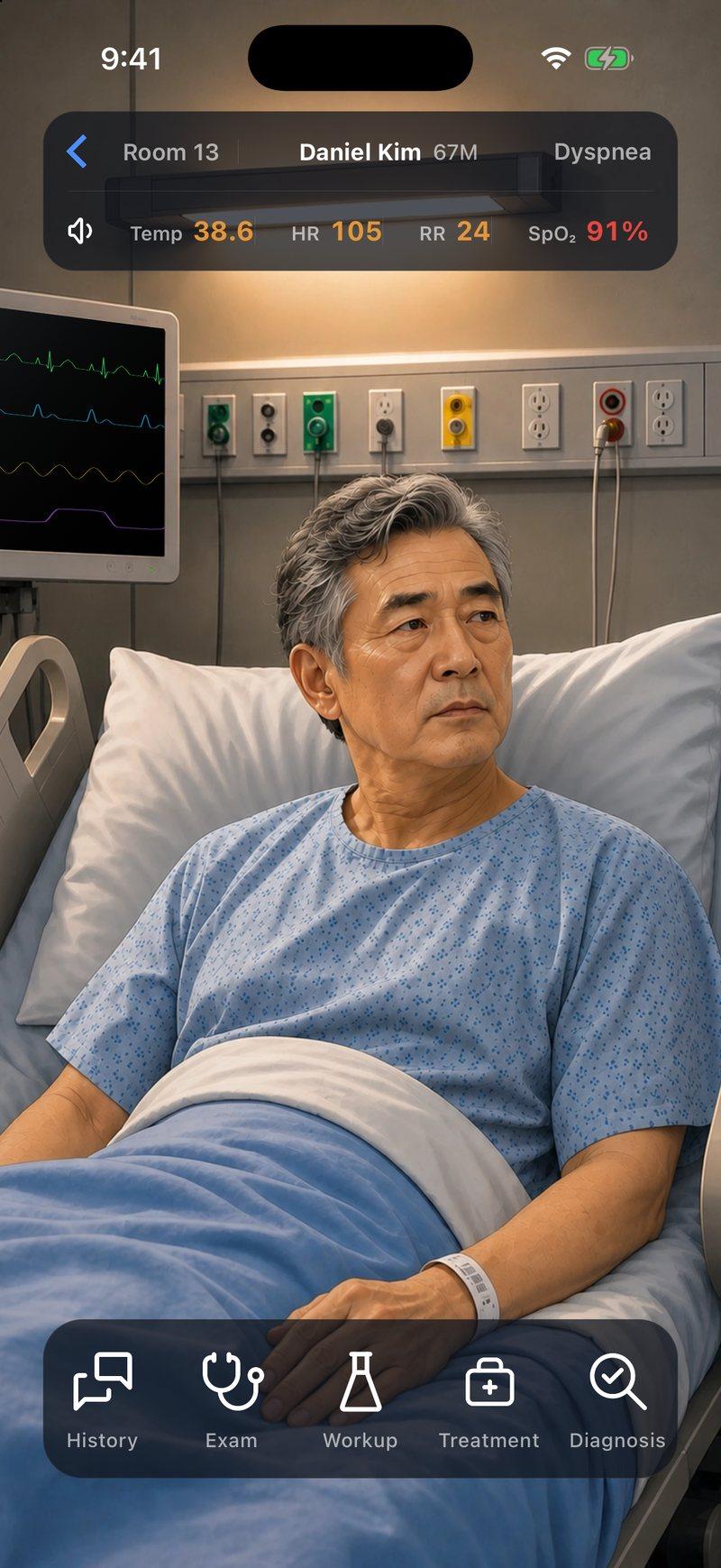
Pneumonia may cause cough, fever, chest pain on breathing in or shortness of breath, with crackles, bronchial breathing or dullness over one area. Not every finding is required. Daniel, 67, had three days of cough and fever. His temperature was 101.5°F (38.6°C), he was breathing 24 times a minute and there were crackles at the base of his right lung. Older adults may instead arrive confused, after a fall or not eating, without a fever. When pneumonia is suspected, consider chest imaging.
Right away
Judge severity at the bedside
Judge severity on the patient’s physiology. Look at the resting oxygen saturation and the work of breathing, the blood pressure and mental state, the urine output and lactate, and whether each is getting better or worse. Daniel’s oxygen saturation was 91% on room air, with a heart rate of 105 and a normal blood pressure.
Two separate questions
Hospital, and then intensive care
The Pneumonia Severity Index uses twenty factors, weighted heavily by age and other illnesses, to estimate the risk of death. It informs where to treat, but it does not decide it. A new oxygen need that persists is a strong reason for hospital care, even with a reassuring score. Intensive care is a separate question. Invasive mechanical ventilation (a breathing tube and a ventilator), or septic shock needing vasopressors (drugs to support blood pressure), means intensive care on its own. Three or more minor criteria identify severe pneumonia and prompt consideration of intensive care. Examples include breathing 30 or more times a minute, poor oxygenation, confusion, pneumonia in several lobes or low blood pressure needing fluids.
Before the patient leaves the department
Give the first antibiotic dose promptly
Cover the common organisms. For patients admitted with pneumonia that is not severe, the usual regimen pairs a beta-lactam, such as ceftriaxone, which covers pneumococcus and Haemophilus, with a macrolide, such as azithromycin, which covers Legionella and Mycoplasma. No bedside finding reliably rules those out. Doxycycline can replace the macrolide when one is unsuitable, and levofloxacin alone is an accepted alternative. A low procalcitonin on its own should not hold back antibiotics in pneumonia that is suspected and seen on the X-ray.
Only with a reason
Broaden for a named resistance risk
Previous respiratory cultures growing MRSA (methicillin-resistant Staphylococcus aureus) or Pseudomonas are important resistance risks. A recent hospital stay with intravenous antibiotics also matters, judged with how sick the patient is and local data. Living in a care facility is not, on its own, a reason for broad coverage. Blood cultures are not routine in pneumonia that is not severe: send them for severe disease or when broadening for resistant organisms. Reassess at about 48 hours, and narrow when results and improvement allow.
Before deciding where
Treat the low oxygen, then reassess
Give oxygen to a target that suits the patient, then check how much is still needed. Going home requires all of these: oxygen levels near the patient’s usual, acceptable work of breathing, stable vital signs, eating and drinking enough, and a plan that works, with an antibiotic the patient can get, follow-up and a way to come back. When breathlessness on walking is the complaint, checking oxygen while walking can show less reserve than the patient has at rest. It is a check for that patient, not a universal rule. Rising oxygen needs, harder breathing, confusion or a falling blood pressure call for early critical care review.
References (7)
- Diagnosis and Treatment of Adults With Community-acquired Pneumonia: An Official Clinical Practice Guideline of the American Thoracic Society and Infectious Diseases Society of America. American Journal of Respiratory and Critical Care Medicine, 2019.
- Diagnosis and Management of Community-acquired Pneumonia: An Official American Thoracic Society Clinical Practice Guideline. American Journal of Respiratory and Critical Care Medicine, 2025.
- Clinical Policy: Critical Issues in the Management of Adult Patients Presenting to the Emergency Department With Community-Acquired Pneumonia. Annals of Emergency Medicine, 2021.
- ERS/ESICM/ESCMID/ALAT Guidelines for the Management of Severe Community-Acquired Pneumonia. Intensive Care Medicine, 2023.
- A Prediction Rule to Identify Low-Risk Patients With Community-Acquired Pneumonia. New England Journal of Medicine, 1997.
- Oxygen Saturations Less Than 92% Are Associated With Major Adverse Events in Outpatients With Pneumonia: A Population-Based Cohort Study. Clinical Infectious Diseases, 2011.
- Procalcitonin as a Marker of Etiology in Adults Hospitalized With Community-Acquired Pneumonia. Clinical Infectious Diseases, 2017.
In the app, Daniel’s Attending’s Notes go deeper: the antibiotic choices, the severity criteria, pitfalls and the evidence, with 11 sources linked to the originals.
Written by Karim Ali, MD, board-certified emergency physician. Reviewed November 2026.
Now make the call yourself
Daniel Kim, 67, has been coughing for three days, has a fever and is getting more short of breath. Work out how sick he is, read his chest X-ray and decide where he should be treated.


Daniel is a fictional patient, and this page is a summary for learning. Code Status is for training only and is not intended for real-world patient care, diagnosis, treatment, or clinical decision-making.