Learn · Cardiology · Beginner
Acute pericarditis
The ECG from a Code Status case, how it differs from a STEMI, and what to do next.
Written by Karim Ali, MD, board-certified emergency physician. Reviewed November 2026.
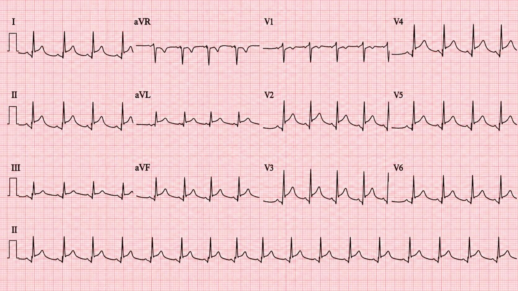
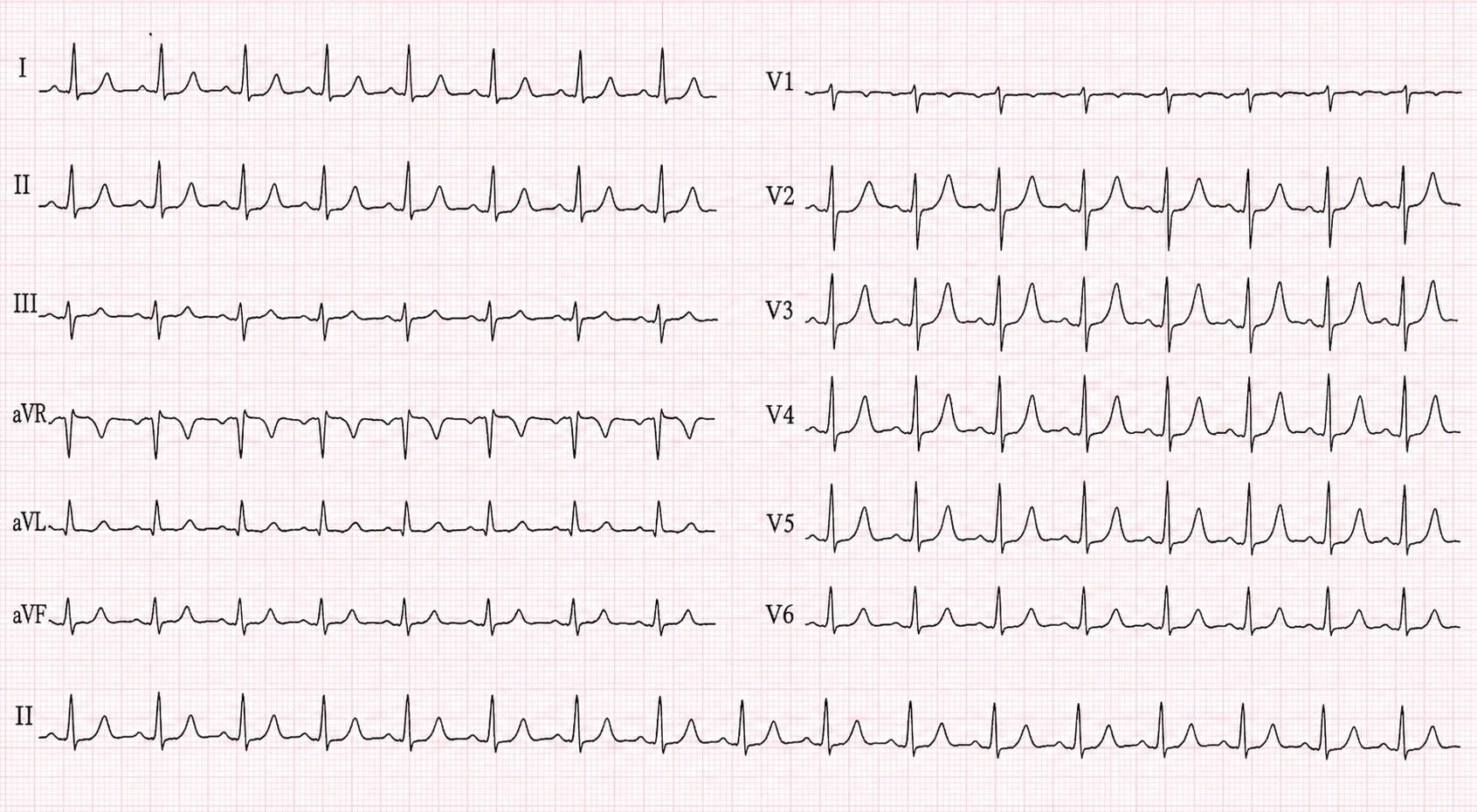
The ST elevation crosses coronary territories: the bottom of the heart (II and aVF), the front (V2 to V4) and the side (I, V5 and V6).
Arjun Gill’s ECG, from his case. Open it full size
Reading this ECG
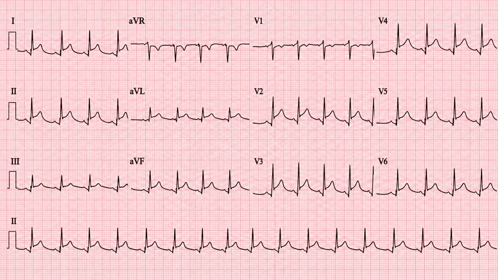
Start with the ST segment, as you would for any chest pain, measured at the J point against the TP segment, the line between the T wave and the next P wave. Here it is raised in I, II, aVF and V2 to V6, about 2.5 mm in lead II, with a concave, scooped shape.
Then look just before the QRS, at the PR segment. In II, aVF and V4 to V6 it sits below the baseline, up to about 1 mm in lead II, deepest just before the QRS. aVR, which looks at the heart from the opposite side, shows the reverse: its ST dips slightly and its PR rises slightly.
That pattern points away from a STEMI, but it does not rule one out. A STEMI usually raises the ST in one coronary territory and pulls it down in the leads facing the opposite wall. Here the elevation crosses territories, and no lead other than aVR shows ST depression. ST depression in any lead other than aVR or V1 should make you think STEMI, and with raised ST in II, III and aVF, ST depression in aVL favors STEMI. Shape alone cannot make the diagnosis, and many patients never show the classic picture, so read it with the story.
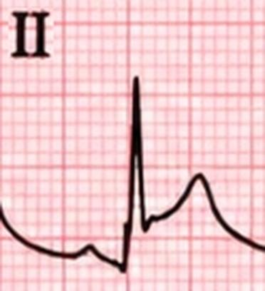
Lead II, enlarged
In the emergency department
From the first ECG to the plan for home, in order.
On arrival
Read the ECG with the story
Widespread ST elevation with PR depression, in someone whose sharp chest pain is worse lying flat and better sitting forward, points to pericarditis. A friction rub supports it, but it is heard in only about a third of patients.
Before you call it pericarditis
Make sure it is not a STEMI
If the tracing could be a STEMI, repeat it, look on echo for part of the heart wall that moves poorly, and show it to cardiology early. Do not wait for a troponin to decide whether a likely STEMI needs the cath lab: early in a heart attack it can still be normal. Arjun’s pain had started 12 hours before, so his normal troponin counted.
The workup
Make the diagnosis and look for complications
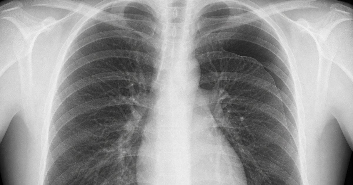
An ECG, a chest X-ray, an echo, a troponin and a C-reactive protein (CRP). Under the 2025 European Society of Cardiology (ESC) guideline, the diagnosis needs the typical pain plus at least two of: a rub, widespread ST elevation or PR depression, a raised CRP, or a new or growing effusion. Arjun had all four: a faint rub, the ECG changes, a raised CRP and a small effusion.
Treatment
An NSAID plus colchicine
An NSAID such as ibuprofen, or aspirin, at full dose until the pain settles and the CRP is normal, then a slow taper. Add colchicine for about 3 months to lower the chance of it coming back, and a proton pump inhibitor to protect the stomach. Steroids are not routine first-line treatment: they are linked to more recurrences.
Before he leaves
Decide who goes home
A fever above 38 °C (100.4 °F), a large effusion, tamponade, or no improvement after about a week of treatment are high-risk features, and these patients are usually admitted. A raised troponin (the heart muscle is involved too), a weakened immune system, trauma or a blood thinner also calls for a closer look. Low-risk patients like Arjun go home with a visit in 1 to 2 weeks, no strenuous exercise until it has fully settled, and for at least a month, and clear reasons to come back, such as breathlessness or lightheadedness.
References (5)
- 2025 ESC Guidelines for the Management of Myocarditis and Pericarditis. European Heart Journal, 2025.
- Evaluation and Treatment of Pericarditis: A Systematic Review. JAMA, 2015.
- A Randomized Trial of Colchicine for Acute Pericarditis (ICAP). New England Journal of Medicine, 2013.
- Colchicine in Addition to Conventional Therapy for Acute Pericarditis: Results of the COPE Trial. Circulation, 2005.
- Indicators of Poor Prognosis of Acute Pericarditis. Circulation, 2007.
In the app, Arjun’s Attending’s Notes go deeper: doses, pitfalls and the evidence, with 10 sources linked to the originals.
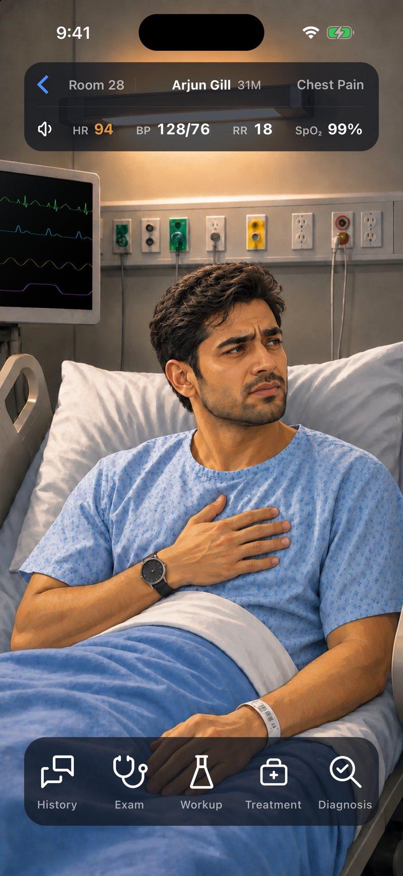
Now make the call yourself
Arjun Gill, 31, arrives with sharp chest pain that is worse when he breathes in. Take the history, examine him, read his ECG and decide what happens next.


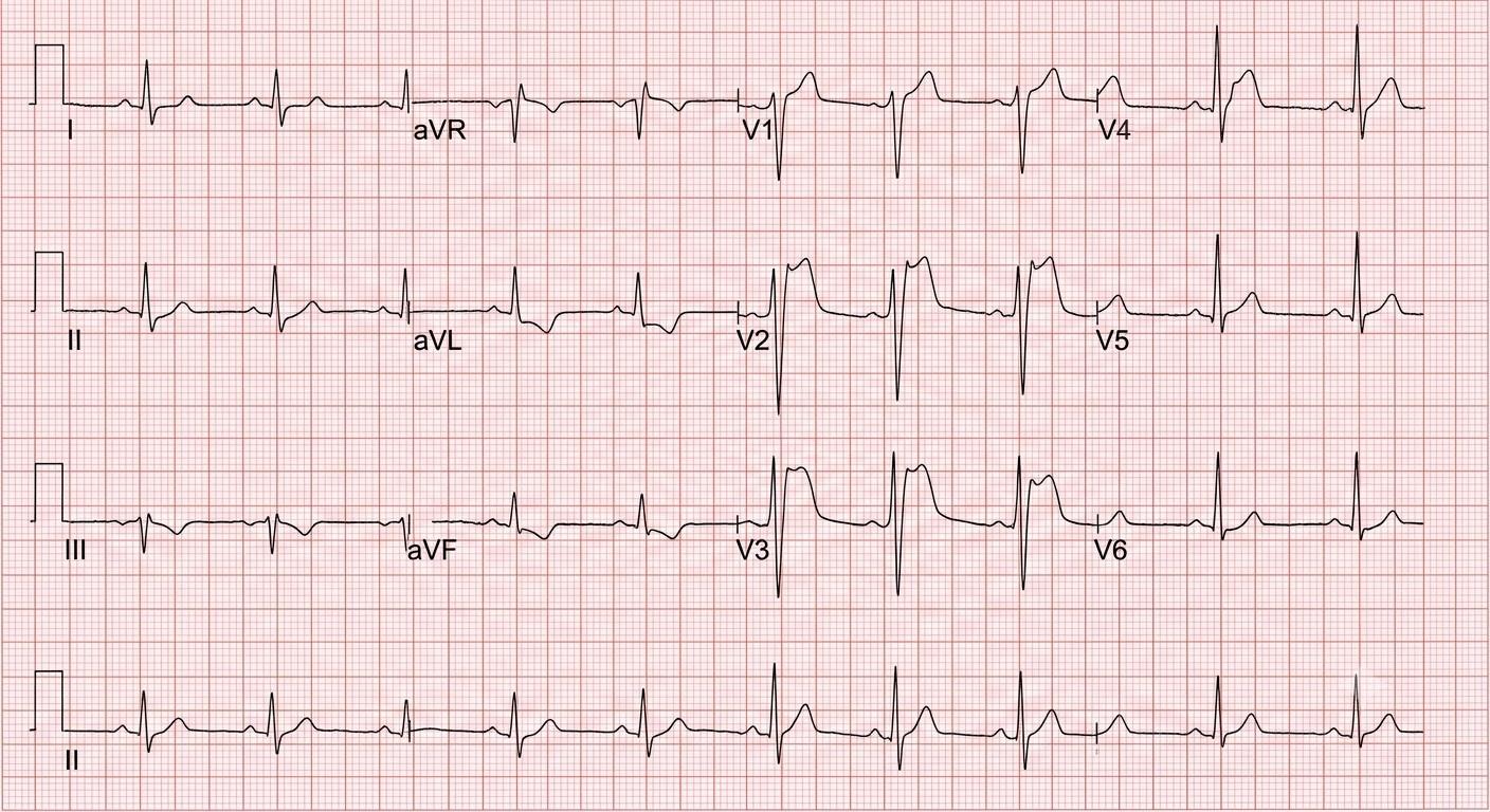
Arjun is a fictional patient, and this page is a summary for learning. Code Status is for training only and is not intended for real-world patient care, diagnosis, treatment, or clinical decision-making.