Learn · Cardiology · Advanced
Acute aortic dissection
The CT angiogram from a Code Status case, and why the heart rate comes down before the blood pressure.
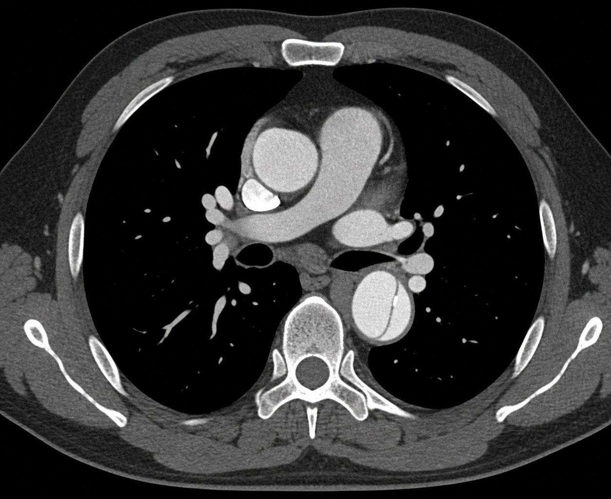
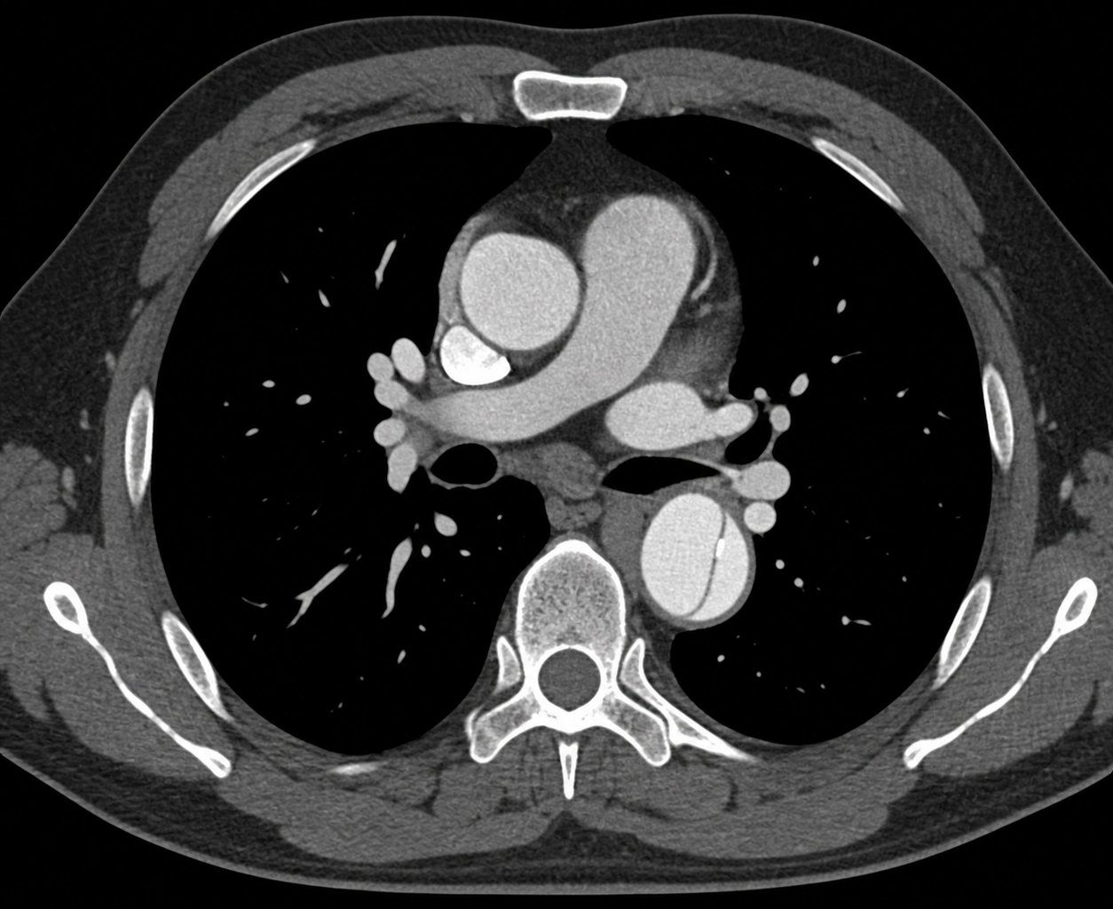
A thin dark line splits the descending aorta, beside the spine, into two channels. The ascending aorta, in front, is clear.
Diane Kessler’s CT angiogram, from her case. Open it full size
Reading this CT
Find the ascending and descending aorta first. The ascending aorta is the round vessel in front, near the middle of the chest. The descending aorta is the round vessel at the back, beside the spine. Contrast makes the blood inside both of them bright.
Here the ascending aorta is evenly bright all the way across. In the descending aorta, a thin dark line crosses the bright contrast and divides it into two channels. That line is the flap. A tear has let blood in between the layers of the aortic wall, so blood now runs in the original channel (the true lumen) and in a false channel inside the wall (the false lumen).
The key question is whether the ascending aorta is involved. If it is, it is Stanford type A. If it is not, it is type B. On this slice the flap is in the descending aorta and the ascending aorta looks clear. That suggests type B, but only the full scan can confirm that the ascending aorta is not involved.
The full scan answers four questions. Is there a dissection? Where does the tear begin? Which branch arteries are involved, and is blood flow to the organs reduced? Has it ruptured? So follow the arteries to the gut, kidneys and legs, and look for leaking contrast, blood around the aorta, or fluid around the lungs or heart.
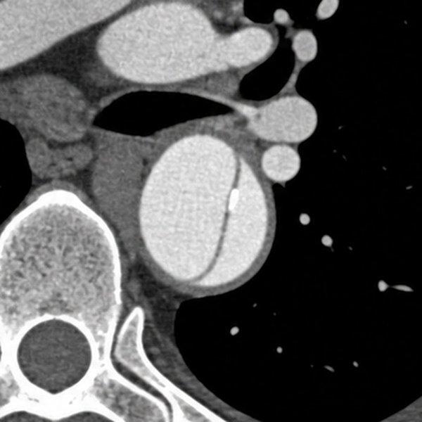
Descending aorta, enlarged
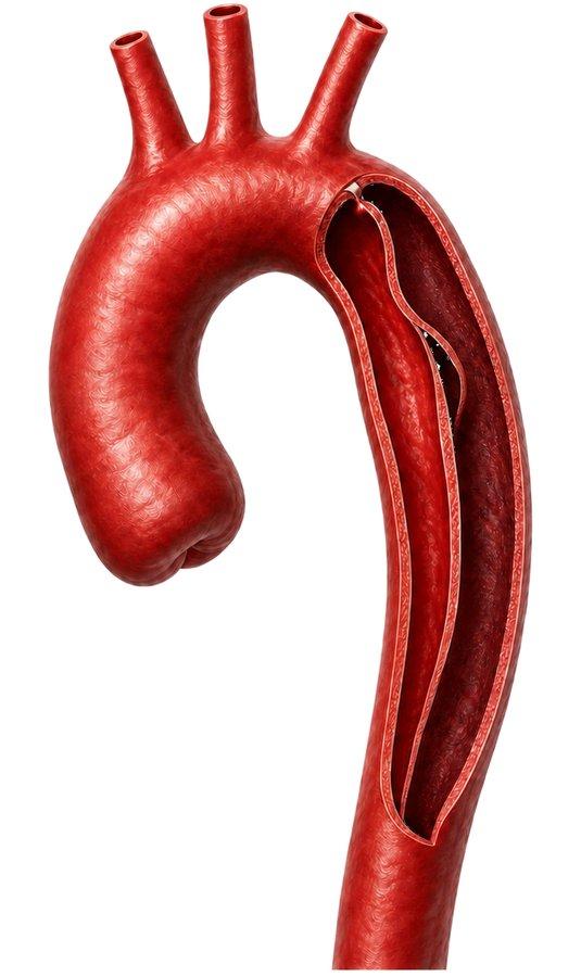
What is happening
The drawing shows the same thing from the outside. The inner lining has torn, and blood has opened a false channel inside the wall of the descending aorta, running down along it.
The flap on the CT is that torn inner layer, seen in a single slice.
In the emergency department
Recognize it, confirm it, slow the heart, then identify the type.
On arrival
Ask how the pain started
Ask whether the pain was at its worst from the very start. Abrupt, severe chest or back pain raises concern for aortic dissection, although other emergencies can start suddenly too. Diane, 62, was doing the dishes when the pain hit all at once. It went from the front of her chest straight through to her back, between her shoulder blades, and the first minute was the worst. High blood pressure is the most common predisposing condition. Hers was 186 over 104, after fifteen years of imperfect control. She drove herself in and looked well. That did not rule it out.
At the bedside
Normal findings do not rule it out
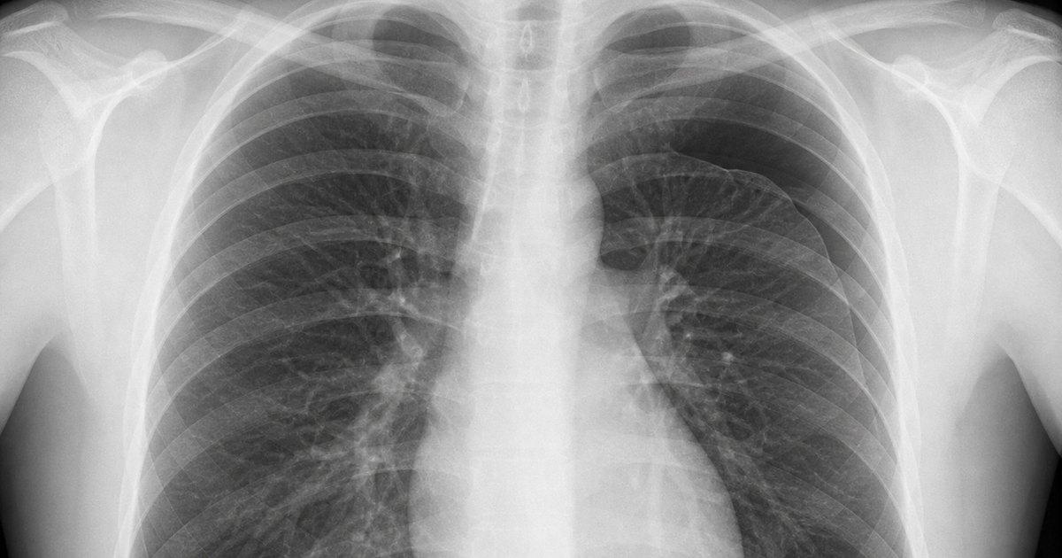
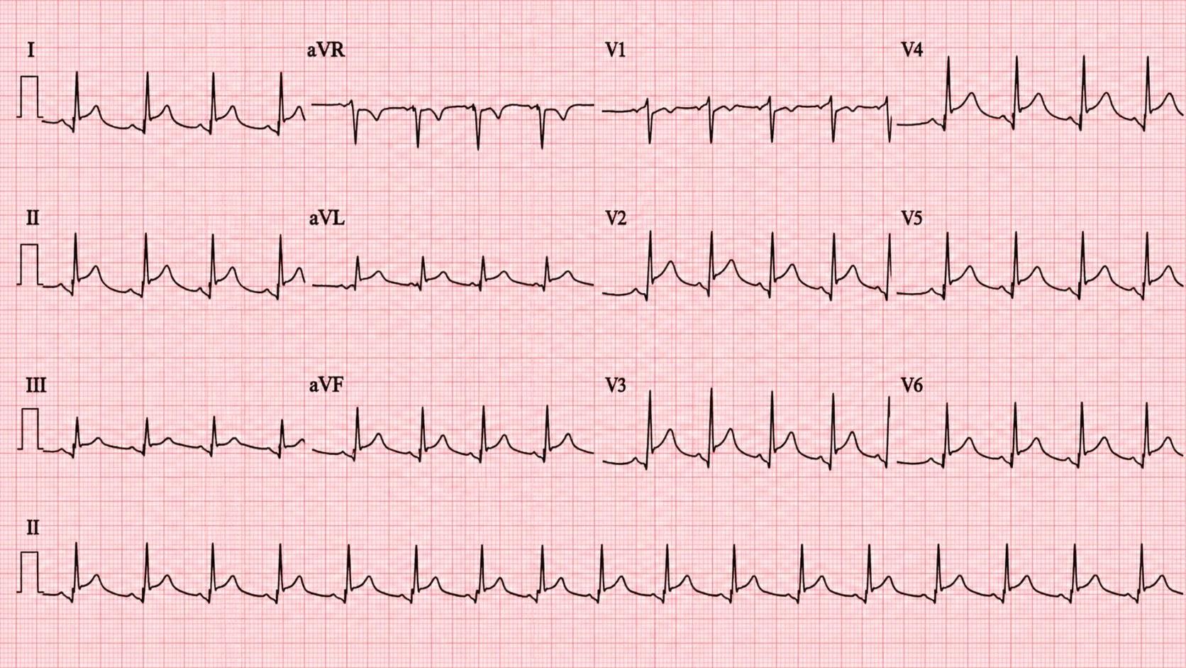
Look for a pulse deficit, a difference in systolic pressure between the arms above about 20 mm Hg, a new diastolic murmur, a new neurologic deficit, a cold pulseless limb or fainting. Most dissections have no pulse deficit and no arm difference, so their absence proves nothing. Diane’s pulses were equal and her arms differed by 4 mm Hg. A normal chest X-ray does not rule it out either: a substantial minority of dissections have a normal film. The ECG and troponin look for a heart cause, and a bedside heart ultrasound can show complications such as fluid around the heart. None of them reliably sees the whole aorta.
When you suspect it
Get a CT angiogram
In a stable patient, CT angiography of the aorta confirms the diagnosis. A high-risk story goes to the scan regardless. A D-dimer is not a stand-alone rule-out. In selected low-risk patients, a score of 0 or 1 on the Aortic Dissection Detection Risk Score together with a negative D-dimer may help avoid the scan. Neither should override a worrying history or examination.
Before blood thinners
Consider dissection first
A type A dissection can involve a coronary artery and look like a heart attack. When dissection is strongly suspected, a clot-dissolving drug can cause catastrophic bleeding. Do not start anticoagulation to treat the dissection itself. If there is a separate reason for it, a specialist weighs the bleeding and clotting risks. None of this is a reason to delay the workup or treatment of a true heart attack.
Once it is confirmed
Heart rate first, then blood pressure
Lower the force of each heartbeat first, with an intravenous beta blocker such as esmolol or labetalol, to a heart rate of about 60 to 80 beats a minute. Then, if the systolic pressure is still above 120 mm Hg, add an intravenous vasodilator such as nicardipine or nitroprusside, aiming under 120 or the lowest pressure that still keeps the organs perfused. Do not give the vasodilator first: it can speed the heart up and add stress on the aortic wall. Treat the pain early, because pain drives the heart rate up. These targets hold only while blood flow to the organs allows: watch alertness, urine output, pulses and lactate, and back off if any worsens. New low blood pressure is a warning, not a success. Pause the infusions and look for rupture, blood around the heart, organs losing blood flow or too much medicine.
Then decide
Type A or type B
Type A involves the ascending aorta and needs emergency cardiothoracic surgery, with rate and pressure control running in the meantime, because untreated mortality climbs by the hour. Uncomplicated type B starts with medicine: intensive monitoring, rate and pressure control, pain relief and repeated examination, with vascular surgery involved from the diagnosis. Type B becomes complicated with rupture, an organ or limb losing its blood supply, pain or blood pressure that will not settle, or an aorta that is widening or a dissection that is extending. That needs urgent vascular review for a repair, usually from inside the artery (endovascular repair) when the anatomy allows.
References (6)
- 2022 ACC/AHA Guideline for the Diagnosis and Management of Aortic Disease. Circulation, 2022.
- EACTS/STS Guidelines for diagnosing and treating acute and chronic syndromes of the aortic organ. European Journal of Cardio-Thoracic Surgery, 2024.
- STS/AATS Clinical Practice Guidelines on the Management of Type B Aortic Dissection. The Annals of Thoracic Surgery, 2022.
- ACR Appropriateness Criteria: Suspected Acute Aortic Syndrome. Journal of the American College of Radiology, 2021.
- Clinical Policy: Critical Issues in the Evaluation and Management of Adult Patients With Suspected Acute Nontraumatic Thoracic Aortic Dissection. Annals of Emergency Medicine, 2015.
- Diagnostic Accuracy of the Aortic Dissection Detection Risk Score Plus D-Dimer for Acute Aortic Syndromes: The ADvISED Prospective Multicenter Study. Circulation, 2018.
In the app, Diane’s Attending’s Notes go deeper: the drug doses, the risk score and the trials on type B repair, with 11 sources linked to the originals.
Written by Karim Ali, MD, board-certified emergency physician. Reviewed November 2026.
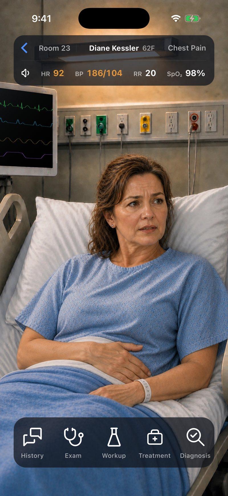
Now make the call yourself
Diane Kessler, 62, arrives with chest pain that came on all at once and went through to her back. She thinks she strained a muscle. Take her history, choose her workup and decide what to give her and where she goes.


Diane is a fictional patient, and this page is a summary for learning. Code Status is for training only and is not intended for real-world patient care, diagnosis, treatment, or clinical decision-making.