Learn · Cardiology · Beginner
Acute decompensated heart failure
The chest X-ray from a Code Status case, and why congestion and blood flow to the organs are two separate questions.
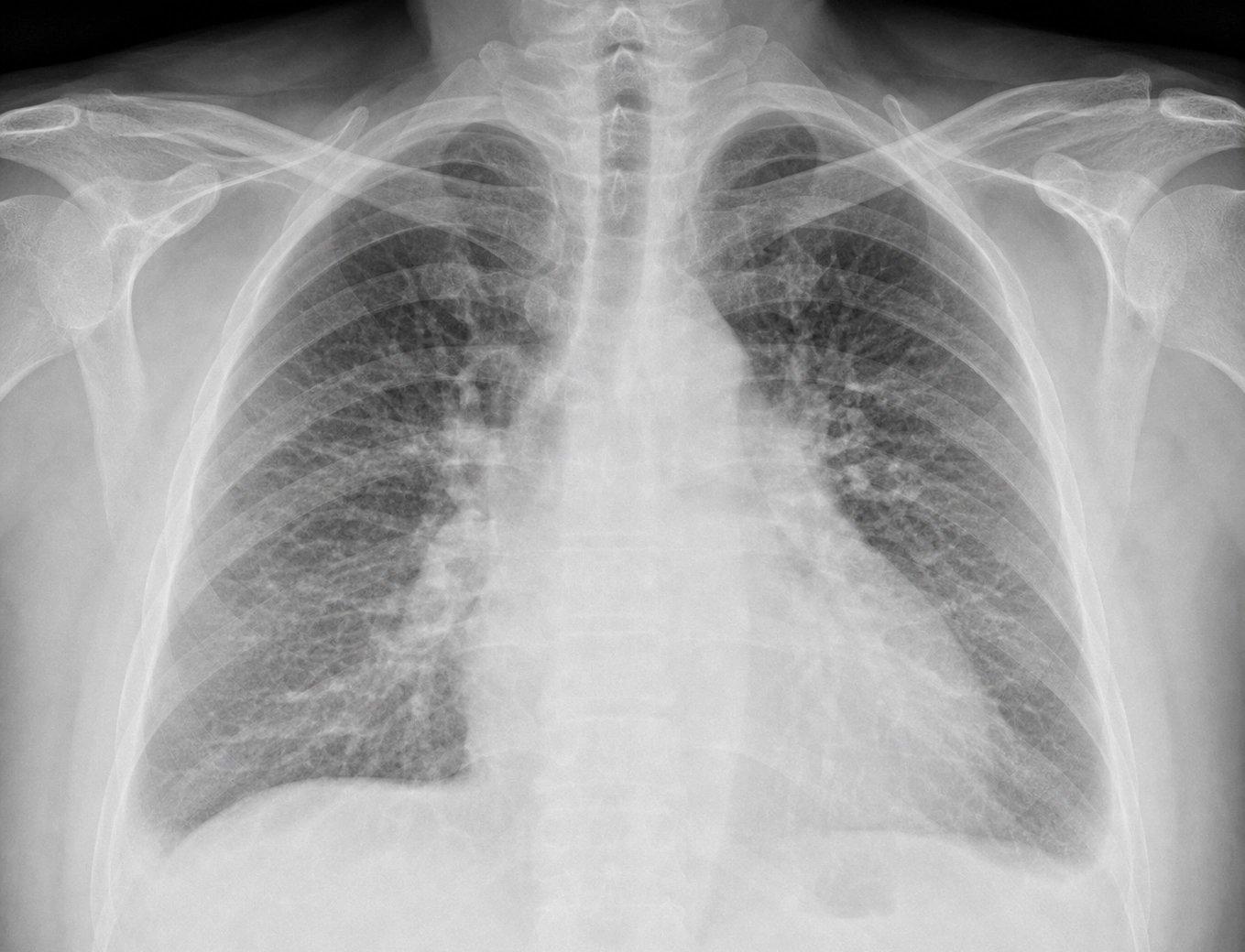
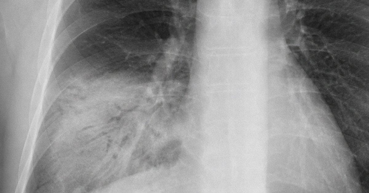
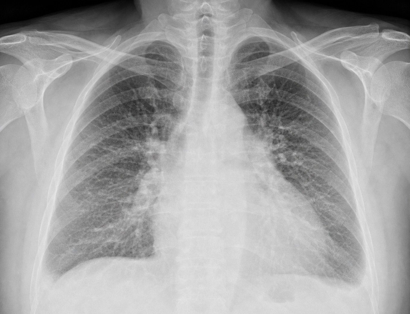
Hazy markings spreading out from the middle of both lungs toward the bases, with a heart that is not enlarged.
Angela Brooks’s chest X-ray, from her case. Open it full size
Reading this X-ray
Start near the middle of each lung, at the hila, where the blood vessels enter. Here the markings look more prominent and hazy than normal, and they spread toward the lower lungs. This pattern can reflect pulmonary vascular congestion and edema: fluid backing up into the lungs. On this film the changes are subtle, so read them with the patient.
The heart is not enlarged. A normal heart size does not rule out acute heart failure.
Other signs include Kerley B lines, fine horizontal lines near the edges of the lungs, and small pleural effusions, which blunt the lower outer corners of the lungs. Neither is clearly seen here. Then look for another cause of the breathlessness, such as pneumonia or a pneumothorax. There is neither.
Lung ultrasound adds another clue. In Angela’s scan, bright vertical lines called B-lines run down from the pleural line. Several across both lungs support fluid in the lung tissue, especially when the examination also suggests congestion. They are not specific to heart failure: pneumonia, acute respiratory distress syndrome and scarred lungs make them too, so where they are and the rest of the picture matter.
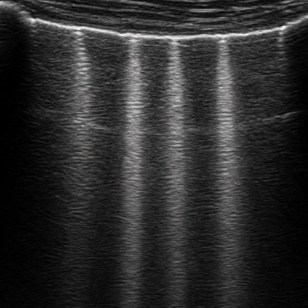
Lung ultrasound, B-lines
In the emergency department
Support the breathing, find the congestion, check blood flow, then treat the cause.
On arrival
Recognize congestion
Angela, 66, has been more and more short of breath for a week. She now sleeps sitting up in a recliner, and some nights she wakes up short of breath. Both suggest fluid backing up into her lungs. On arrival her oxygen saturation was 88% on room air and her blood pressure 158/92. Look for crackles, full neck veins and leg swelling. Pulmonary edema can also wheeze, so a wheeze does not always mean asthma.
At the same time
Check blood flow to the organs
Congestion and poor blood flow are different problems. A patient can have fluid-filled lungs and still be warm, alert and well perfused. Cool hands and feet, confusion, less urine or low blood pressure raise concern that the circulation is failing. The classic split is warm and wet versus cold and wet. A cold, poorly perfused patient needs urgent assessment for cardiogenic shock, including a bedside heart ultrasound, even when the blood pressure is not very low.
The workup
Confirm the picture and find the trigger
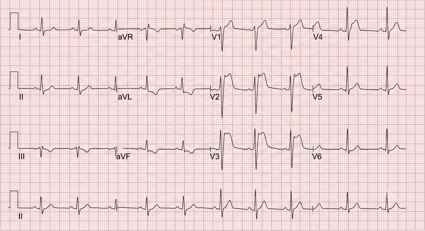
The chest X-ray and lung ultrasound support the diagnosis. BNP or proBNP is most useful when the cause of the breathlessness is uncertain: a low value makes acute heart failure much less likely, though the result depends on the setting. An ECG, troponin, kidney function and electrolytes look for triggers and complications. A raised troponin can mean heart muscle injury without a heart attack.
For the breathing
Support the breathing
Give oxygen when the saturation is low. For marked breathing distress or oxygen levels that stay low, noninvasive ventilation (a tight mask that pushes air in under pressure) can improve oxygen levels and reduce the work of breathing, if the patient is suitable. When a sharp rise in blood pressure has pushed fluid into the lungs, nitroglycerin can quickly lower the heart’s filling pressures and ease the breathing. Avoid it with low blood pressure, a recent erectile dysfunction medicine or a suspected right ventricular infarction. Do not wait for every test result before supporting the breathing.
To remove the fluid
An IV diuretic, not IV fluids
For a patient with excess fluid, a loop diuretic such as furosemide, given into a vein, is the core treatment. The starting dose depends on what the patient already takes. Then follow the urine output, breathing, blood pressure, kidney function and electrolytes, and adjust to the response. Avoid routine IV fluids: a bolus raises filling pressures that are already too high. Not every patient with pulmonary edema has much extra fluid in the body, so match the treatment to the patient.
Before choosing a bed
Reassess breathing and blood flow
Better oxygen levels do not prove better blood flow. Recheck the breathing, oxygen needs, blood pressure, alertness and signs of congestion or poor blood flow. Breathing failure, shock or worsening organ function needs intensive care and specialists. A patient who is stable but still needs oxygen, IV treatment or close watching needs a ward bed. Home only when hospital-level care is no longer needed.
References (6)
- 2022 AHA/ACC/HFSA Guideline for the Management of Heart Failure. Circulation, 2022.
- 2026 ESC Guidelines for the Management of Heart Failure. European Heart Journal, 2026.
- Lung Ultrasound in Acute and Chronic Heart Failure: A Clinical Consensus Statement of the European Association of Cardiovascular Imaging. European Heart Journal: Cardiovascular Imaging, 2023.
- Heart Failure Association of the ESC Practical Guidance on the Use of Natriuretic Peptide Concentrations. European Journal of Heart Failure, 2019.
- Fifth Universal Definition of Myocardial Infarction (2026). Journal of the American College of Cardiology, 2026.
- 2025 Concise Clinical Guidance: An ACC Expert Consensus Statement on the Evaluation and Management of Cardiogenic Shock. Journal of the American College of Cardiology, 2025.
In the app, Angela’s Attending’s Notes go deeper: the doses, the pitfalls and the evidence, with 11 sources linked to the originals.
Written by Karim Ali, MD, board-certified emergency physician. Reviewed November 2026.
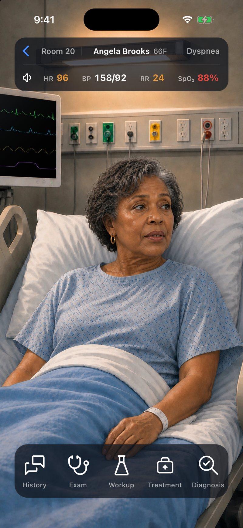
Now make the call yourself
Angela Brooks, 66, has been more and more short of breath for a week and can no longer lie flat to sleep. Her oxygen saturation is 88% on room air. Assess her breathing and circulation, read her imaging and decide her treatment and where she goes.


Angela is a fictional patient, and this page is a summary for learning. Code Status is for training only and is not intended for real-world patient care, diagnosis, treatment, or clinical decision-making.